
Since the year 1974, the Nikon Small World Photomicrography Competition has helped to highlight the inner artists in scientists, researchers, and doctors. A whole new world was opened up to the public as they used their skills and creativity combined to capture life. It is safe to say that this year’s contest was as thrilling. The winners were a group of scientists from the National Institute of Health.
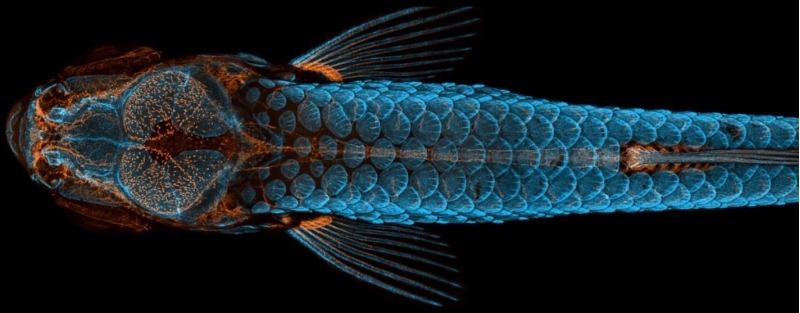
Daniel Castranova, who was assisted by Bakary Samasa along with Dr. Brant Weinstein happened to capture the winning image. It was of a young zebrafish.
“The image is beautiful, but also shows how powerful the zebrafish can be as a model for the development of lymphatic vessels,” Castranova said, “Until now, we thought this type of lymphatic system associated with the nervous system only occurred in mammals. By studying them, the scientific community can expedite a range of research and clinical innovations–everything from drug trials to cancer treatments. This is because fish are so much easier to raise and image than mammals.”
Life under a microscope is so colorful and beautiful.
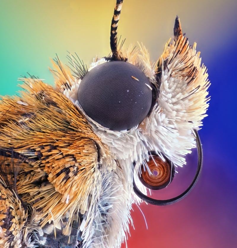

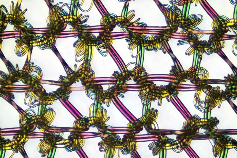
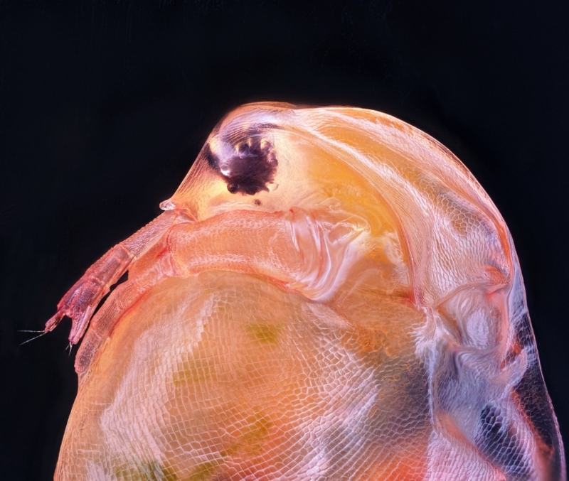
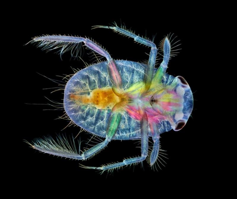
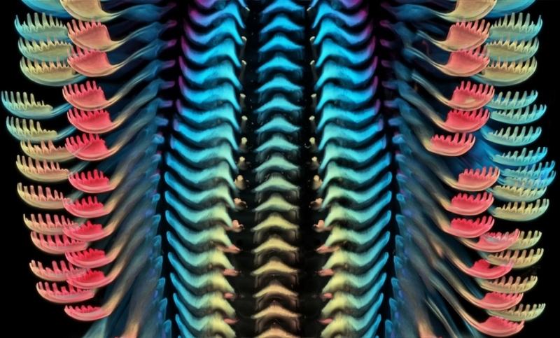

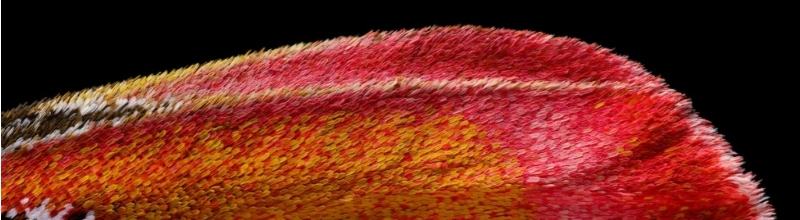
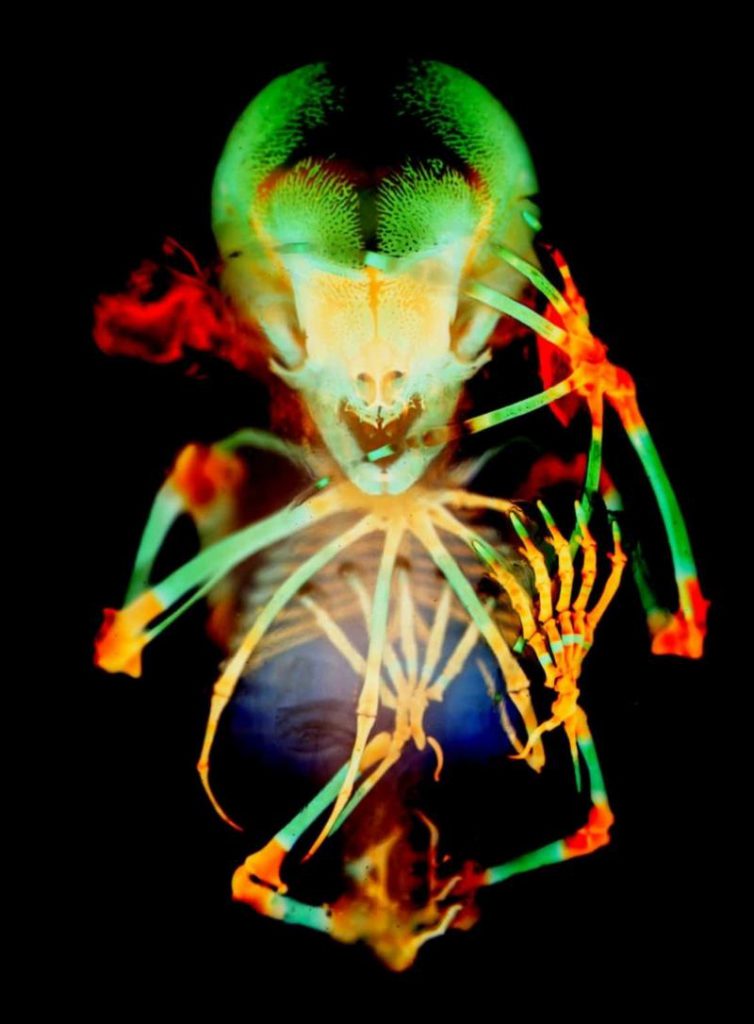
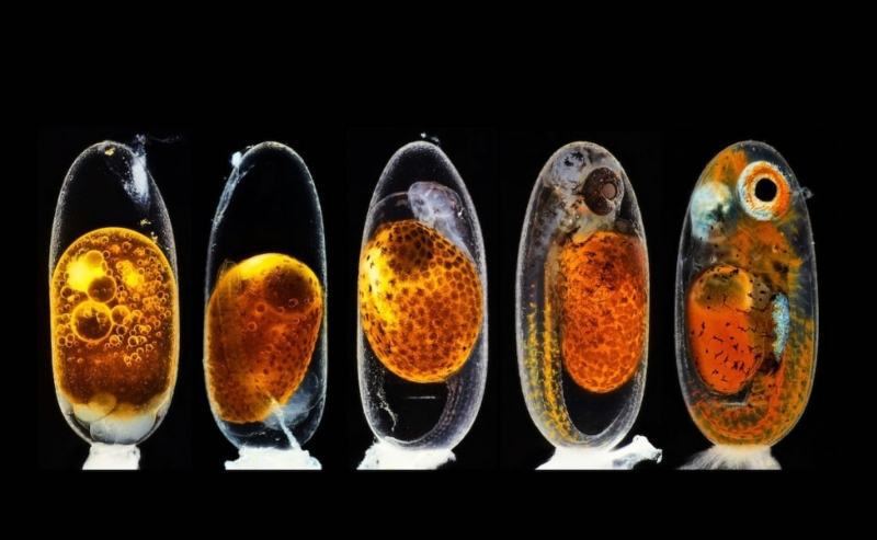
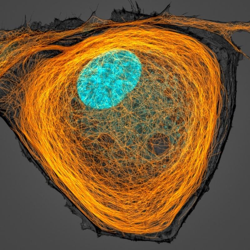

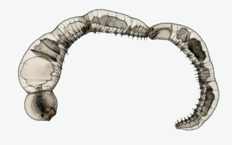
Image Source – Nikon Small World: Facebook | Instagram
There were over 2,000 pictures that were entered into this year’s competition and you just took a look at the top photographs. Head over to Nikon Small World website to see when this year’s winners will be released. Life under this microscopic world is a real marvel.
Related Articles:
South Dakota Sculptor Recycles Old Farm Equipment Into Extraordinary Animal Sculptures
The ‘Under-Dog’ Project Of Canine Pictures From This Angle Will Make Your Day
Artist Uses A Chainsaw And Transforms A Fallen Redwood Tree Into A Giant Octopus